Edit Content


As of July 2025, seven people have been implanted with Neuralink’s brain-computer interface (BCI). The implant enables individuals with cervical spinal cord injuries or amyotrophic lateral sclerosis (ALS) to control a computer with their thoughts. Could this endeavor mark a step toward “biocomputers”? Could it provide deeper insights into how the brain functions and how it is affected by neurodegenerative
conditions?
More importantly, how does Hawai‘i adapt to this ever-evolving landscape of neuroscience innovations
and developments?
We are addressing this challenge through Hawai‘i BRAIN (Bridging Research & Innovation in Neuroscience), a hybrid research ecosystem that leverages the innovative powerhouse of our global research partners and government agencies to support high-impact neuroscience research in Hawai‘i. For over a decade, BRAIN has driven and led Hawai‘i’s Neuroscience RIDE (Research, Innovation, Discovery & Education).
We are meeting this golden age of neuroscience discovery with excitement, passion, continuous
learning, and adaptability, looking ahead to make wise and strategic investments to achieve what we can only accomplish by working laulima together!
Mahalo to our patients and their ‘ohana, who continue to inspire us and are the reason we do what we do every day; our amazing community, who have trusted us to bring the most advanced neuroscience options to the islands; and our global partners, who have collaborated closely with us to attract groundbreaking research opportunities and much-needed healthcare resources (over $3 million and more than 50 healthcare positions last year) to serve our precious local ‘ohana.
We are grateful to University of Hawai‘i John A. Burns School of Medicine Dean Sam Shomaker, MD, for supporting our neuroscience community, especially our students, who are the next generation of physician leaders and scientists—and our hope! We know you will find our speakers’ lectures illuminating and the research work of our students, residents, and faculty
inspiring. Enjoy!
Aloha,
Neuroscience Chair, Hawaii Pacific Neuroscience
Clinical Professor of Med (Neurology), Graduate Faculty, Clinical & Translational
Research
University of Hawai`i John Burns School of Medicine
Aloha, attendees of the 16th Hawai‘i Pacific Neuroscience Symposium,
This year’s theme of The Golden Age of Neuroscience Discovery is a title that feels especially fitting as our world continues to evolve with breathtaking speed in technological advancements.
Today, we are proud to showcase more than 20 research abstracts, with the majority coming from our very own University of Hawai‘i John A. Burns School of Medicine. Your participation here, and the work you present, demonstrate that JABSOM is truly at the forefront of cutting-edge research.
I want to extend a heartfelt mahalo to Dr. Kore Liow for his many years of mentorship. Through the BRAIN program—Bridging Research and Innovation in Neuroscience—he has inspired and guided so many of our students, helping them navigate their research journeys and achieve remarkable accomplishments. Mahalo to all of you for being part of this exciting chapter in neuroscience- and for continuing to push the boundaries of discovery.

Professor and Dean,
University of Hawai`i John Burns School of Medicine
 Research, Innovation, Discovery & Education
www.hawaiineuroscience.com/research/brain/
Hawaii BRAIN (Bridging Research & Innovation in Neuroscience) is a hybrid research ecosystem that leverages the innovative powerhouse of our global research partners and government agencies to support high impact neuroscience research in Hawaii and beyond.
Our primary mission is to turn data driven discoveries through basic
science, translational and clinical research to fuel hope and contribute to the scientific knowledge and advance progress in the fight against neurological diseases especially those affecting Hawaii and the Pacific Islands. The BRAIN is supported by the Hawaii Pacific Neuroscience Foundation in Honolulu, Hawaii. The BRAIN is responsible for driving and leading Hawaii’s Neuroscience RIDE (Research, Innovation, Discovery & Education) for over a decade now.
BRAIN’s Clinical Research Center is recognized nationally and on the forefront of working with some of the best biomedical organizations and agencies and in the world including NIH in scientific advancement and development of innovative treatments for some of the most challenging neurological diseases of our times.
At any one time, BRAIN investigators are collaborating with global partners and agencies on more than
30-40 research projects investigating Alzheimer’s, Parkinson’s, MS, Neuromuscular, Epilepsy,
BRAIN investigators are partnering with both private and governmental agencies as part of global collaborative efforts and initiatives to advance knowledge and understanding of etiologies, pathogenesis, pathophysiology of neurological conditions including sampling of biomarkers (CSF, serum, brain tissues), genetic or Imaging data working with NIH, NINDS NeuroCOVID databank, ALZ-NET, Global Alzheimer’s Platform Foundation & others collaborating global partners. One such BRAIN robust research and academic programs focus on use of AI to advance neuroscience at our NeuroAI Research Lab/Alzheimer’s Neural Network EEG (ANNE) Lab investigating the use of AI in Neural Engineering especially as it applies to measuring electrical activities in the brain cortical regions using EEG (Electroencephalogram) collaborating with AI innovators.
BRAIN investigators are partnering with both private and governmental agencies as part of global collaborative efforts and initiatives to advance knowledge and understanding of etiologies, pathogenesis, pathophysiology of neurological conditions including sampling of biomarkers (CSF, serum, brain tissues), genetic or Imaging data working with NIH, NINDS NeuroCOVID databank, ALZ-NET, Global Alzheimer’s Platform Foundation & others collaborating global partners. One such BRAIN robust research and academic programs focus on use of AI to advance neuroscience at our NeuroAI Research Lab/Alzheimer’s Neural Network EEG (ANNE) Lab investigating the use of AI in Neural Engineering especially as it applies to measuring electrical activities in the brain cortical regions using EEG (Electroencephalogram) collaborating with AI innovators.
BRAIN investigators in Center for Rare Neurological Disorders awarded competitive research award funding from n-Lorem to collaborate on a translational research project in the world to assist with trial conception, design, development and conduct of ASO clinical trial on rare genetic disease including writing and submitting protocols for submission to FDA for IND (Initial New Drug) application for single center single participant FDA submission for patient #2 in the world.
BRAIN Scholar Program is part of the University of Hawaii John Burns School of Medicine MD5 MED 599 Neuroscience Research Course. University Hawaii medical students may sign up for elective credit while working at BRAIN in MD5 MED 599 Neuroscience research credit. All BRAIN’s investigators are involved in mentoring research residents and medical students in the BRAIN Scholar program. Every year, over 50 residents, medical, graduate and undergraduate students accepted into Hawaii BRAIN scholar and Intern program mentored by BRAIN investigators.
BRAIN Scholar Program is part of the University of Hawaii John Burns School of Medicine MD5 MED 599 Neuroscience Research Course. University Hawaii medical students may sign up for elective credit while working at BRAIN in MD5 MED 599 Neuroscience research credit. All BRAIN’s investigators are involved in mentoring research residents and medical students in the BRAIN Scholar program. Every year, over 50 residents, medical, graduate and undergraduate students accepted into Hawaii BRAIN scholar and Intern program mentored by BRAIN investigators.

Hawaii Pacific Neuroscience Chair & Clinical Professor of Medicine
(Neurology), Clinical & Translational Research, University of Hawaii
John Burns School of Medicine
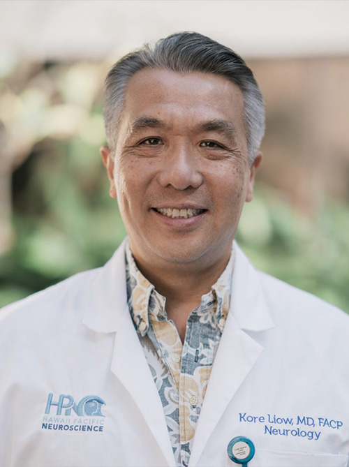
Dr. Kore Kai Liow, a distinguished neurologist, entrepreneur, and CEO of Hawaii Pacific Neuroscience, where he also serves as Neuroscience Chair. As a Clinical Professor of Medicine (Neurology) and Graduate Faculty in Clinical & Translational Research at the University of Hawaii John A. Burns School of Medicine, he is a nationally recognized and sought after key opinion leader and served on committees and advisory panels for NIH and CDC. Trained as a research neurologist at NINDS, NIH, Dr. Liow continues to serve on NIH’s Review Study Section and Grant Review Committee. He spends majority of his time in research and served as principal investigator for over 200 clinical trials, authored over 100 peer-reviewed PubMed publications. He currently oversees the BRAIN program which is mentoring over 20 medical students at UH.. A pragmatic leader, Dr. Liow views challenges as opportunities, emphasizing problem-solving and innovation in advancing neuroscience and patient care.
In His Spare Time: he enjoys Surfing, spending time with his wife ❤️and family and continues to teach Sunday school in his church every week for the last 35 years!

Associate Professor Psychiatry & Behavioral Sciences, Director, Brain
Stimulation Laboratory, Director, Interventional Psychiatry Clinical Research,
Department of Psychiatry and Behavioral Sciences,
Stanford University School of Medicine
Dr. Nolan Williams, Professor of Psychiatry and Behavioral Sciences at Stanford University and Director of the Stanford Brain Stimulation Lab, is a triple board-certified neuropsychiatrist in general neurology, general psychiatry, and behavioral neurology & neuropsychiatry. Renowned for his pioneering work in neuromodulation and rapid-acting therapies, Dr. Williams led the development of Stanford Accelerated Intelligent Neuromodulation Therapy (SAINT), the world’s first non-invasive, rapid-acting treatment for treatment-resistant depression, which received FDA Clearance in 2022 and is now Medicare-reimbursed and widely used globally. His research also explores psychedelic medicines, including groundbreaking trials on ibogaine’s neurobiological effects, and advances neuroimaging-based approaches to optimize therapeutic outcomes for mood disorders, OCD, and other neuropsychiatric conditions.
In His Spare Time: Dr. Williams loves to kite surf in Maui

Director, Headache & Facial Pain Center, Clinical Assistant Professor of
Medicine (Neurology), University of Hawaii John Burns School of Medicine
Dr. Eonjung Angeline Kim, a distinguished headache medicine specialist from the Montefiore Headache Center in New York. Trained in neurology at the Icahn School of Medicine at Mount Sinai and a graduate of the Lewis Katz School of Medicine at Temple University, Dr. Kim is passionate about addressing headache disorders through a comprehensive, patient-centered approach. As the Director of the Headache & Facial Pain Center at Hawaii Pacific Neuroscience, she is dedicated to breaking down socioeconomic barriers to healthcare and serving diverse, underserved communities. Dr. Kim is excited to lead a unique headache program, collaborating with specialists in sleep and preventive lifestyle to advance treatment and research, ultimately enhancing patients’ quality of life. Join us in welcoming Dr. Kim as she shares her expertise and vision for transforming headache care.
In Her Spare Time: Outside of medicine, she enjoys traveling, exploring new foods, and has recently taken up CrossFit.”

Director –MS & Neuroimmunology Center, ALS and Neuromuscular EMG
Center, IV Infusion Ctr Clinical Assistant Professor of Medicine (Neurology),
University of Hawaii John Burns School of Medicine
Dr. Gonzalez, a pioneering neurologist with dual fellowship training in Neuroimmunology and Neuromuscular Disease. Originally from Asturias, Spain, she earned her MD from the University of Oviedo and completed advanced training at prestigious institutions, including Johns Hopkins, Northwestern, and UC Irvine. As the first specialist in Hawaii with this unique expertise, Dr. Gonzalez is leading the establishment of the state’s first MS and Neuromuscular program focused on Neuroimmunology. Her work brings cutting-edge treatments, clinical trials, and research to Hawaii and the Pacific Region, offering new hope for patients with complex neurological conditions.
In Her Spare Time: Dr. Gonzales likes to write books and uses an alias name “Nour Lorenzo”which are written in Spanish and is working on getting the books translated into English! She is also a lover of the outdoors and likes to surf.

Presidential Distinguished Chair & Residency Program, Professor and Director,
Dept. Neurosurgery, Houston Methodist Hospital
Dr. David Baskin, a distinguished neurosurgeon and researcher. Dr. Baskin serves as Professor and Vice Chairman of Neurosurgery at the Methodist Hospital Neurological Institute, where he also directs the Peak Brain and Pituitary Center and the Neurosurgery Residency Program. A graduate of Swarthmore College and Mt. Sinai School of Medicine, he completed his neurosurgical training at the University of California, San Francisco.
Dr. Baskin is a pioneer in pituitary and skull base surgery, having performed over 5,000 endoscopic endonasal operations, and has operated on more than 10,000 patients. His groundbreaking research focuses on brain and spinal cord injury protection, novel brain tumor therapies, and oncomagnetics, a field he helped establish. He is a co-inventor of a device using oscillating magnetic fields to treat cancer, currently in clinical trials. With over $28 million in research funding and 185 publications, including several books, Dr. Baskin is a recipient of numerous awards, including those from the American Association of Neurological Surgeons and the Congress of Neurosurgery. He is listed in Who’s Who in America and consistently recognized among the Best Doctors in America.
In His Spare Time: Dr. Baskin has been in love with Hawaii since his first visit in 1978 when he was an intern in surgery, and has been coming at least once or twice a year ever since. He loves the feel of the sand on his feet, watching the crabs run their intricate patterns, and is a great fan of shave ice and owns his own shave ice machine.

Clinical Lead & Director, Sleep and Insomnia Center, Clinical Assistant Professor of
Medicine U of Hawaii
Dr. Nicholas Anderson, MD, a distinguished Sleep Medicine physician specializing in sleep apnea, insomnia, restless leg syndrome, and pediatric sleep disorders. Trained at the University of Colorado School of Medicine, he completed his Family Medicine residency at the University of Hawaii and a Sleep Medicine fellowship at the University of Utah. Dr. Anderson is dedicated to improving patients’ quality of life through comprehensive, patient-centered care, emphasizing the critical role of sleep in health. An advocate for sleep disorder awareness, he is committed to advancing medical education and public understanding of sleep’s impact on chronic disease management.
In his spare time, he is an avid Denver sports fan and enjoys playing sports including basketball and tennis, spending time outdoors with his wife and children, and participating in his church.

Director, Comprehensive Epilepsy Ctr Director, Video-EEG Epilepsy Monitoring
Unit, Clinical Assistant Professor of Medicine (Neurology), University of Hawaii
John Burns School of Medicine
Dr. DuGas, a distinguished neurologist and director of the Video-EEG Epilepsy Monitoring Unit and EEG Diagnostic Laboratory at Hawaii Pacific Neuroscience. Originally from Vancouver, Canada, Dr. DuGas transitioned from a career in pharmacy to pursue advanced training in neurology, epilepsy, and EEG at Yale Medical School. Under his leadership, the EEG laboratory became the first ABRET-accredited epilepsy center in Hawaii. As co-director of the Comprehensive Epilepsy Center, he is dedicated to delivering evidence-based treatments and advancing cutting-edge research to improve patient outcomes for seizure-related disorders, headache, Parkinson’s Disease, and other neurologic conditions. Dr. DuGas is committed to bringing innovative therapies to Hawaii, transforming lives through his expertise and passion for neuroscience.
In His Spare Time: completed the three levels of the Chartered Financial Analyst program and am studying the history of financial markets.

Behavioral Neurology Fellow,
Cedars-Sinai UCLA
Dr. Michael Sonson is a fellow of behavioral neurology, graduate of the NYU school of medicine, and graduate of the NYU Neurology residency program. This year he will be finishing his training at the Jona Goldrich Center for Alzheimer’s and Memory Disorders at Cedars Sinai Hospital in Los Angeles. His topics of expertise include cognition, memory, personality, and healthy aging and he’s best known for his blend of expertise and compassionate patient care. His research explores the intersection of culture, language, and cognition with efforts to reach and provide for underserved and understudied populations. Today, Dr. Sonson will share his insights on the latest advances in the understanding and treatment of Alzheimer’s disease: innovative developments which will make for patient-centered solutions to a previously unsolvable condition.
In His Spare Time: Michael just recently picked up learning and playing guitar

Professor and Dean, University of Hawaii John Burns School of Medicine.
Dr. Thomas Samuel aka “Sam” Shomaker, a distinguished 1986 graduate of the John A. Burns School of Medicine (JABSOM). Dr. Shomaker served as JABSOM’s vice dean from 2000 to 2005 and interim dean from 2005 to 2006. He later held prominent roles as dean of the University of Texas Medical Branch’s Austin campus and dean and vice president at Texas A&M Health Science Center.
With 20 years of health care experience, Dr. Shomaker holds an MD from UH Mānoa, a JD from Georgetown University School of Law, a master’s in management from Stanford University Graduate School of Business, and a bachelor’s degree from St. Louis University. His extensive expertise and leadership make him an inspiring speaker for our conference.
One of the high lights of Dr. Shomaker’s professional career is the honor of being named the dean of his alma mater John A Burns School of Medicine. He proudly states that he is appreciative of this immense responsibility and thanks you for the opportunity to serve you and the people of the state of Hawaiʻi.


Joshua Grube,1 Dariann Davis,2 Riley Regan,2 Isabella Ford,2 Joshua Wung,2 Janette Bow-Keola,1 Tyrone Sumibcay,1 Matthew Kao,1 Jonathan Phisayavong,3 Amir Meghdadi,4 Chris Berka,4 Enrique Carazzana,1 Kore Kai Liow1,2
1 John A. Burns School of Medicine, University of Hawaiʻi, Honolulu, HI
2 Alzheimer’s Neural Network EEG Research Laboratory, Hawaii Pacific Neuroscience, Honolulu, HI
3 JABSOM Biostatistics Core Facility, Department of Quantitative Health Sciences, University of Hawaiʻi
4 Advanced Brain Monitoring, Carlsbad, CA
Existing research has established an association between hypertension (HTN), mild cognitive impairment (MCI), and Alzheimer’s Disease (AD), and how uncontrolled HTN can increase the progression of MCI to AD. Brain Electrical Activity Mapping (BEAM) has been used to identify unique biomarkers useful for MCI diagnosis and other neurocognitive diseases. This project’s objective was to identify differences between BEAM biomarkers in patients with MCI only to those with a comorbid diagnosis of HTN. A retrospective chart review of patients with an MCI diagnosis who underwent BEAM testing was conducted at Hawaii Pacific Neuroscience. Additional criteria for inclusion were a history of HTN, no stroke or TBI within 2 years prior to BEAM testing, no concurrent antidepressant/antipsychotic medication use, and no diagnosis of other vascular diseases, such as Type II DM and various vasculitides. Initial analysis revealed no significant differences between MCI only (N=42) vs. MCI with HTN (N=11) groups for patients stratified by MMSE scores (25-30 for Group 1 [N=28] and 20-24 for Group 2 [N=18]) for Auditory Oddball (AO) N1 Peak Latency, AO P300 Maximum Amplitude, and AO P300 Maximum Latency. Further data collection will increase sample size/power, and other disease biomarkers may be explored.
1 John A. Burns School of Medicine, University of Hawaiʻi, Honolulu, HI
2 Alzheimer’s Neural Network EEG Research Laboratory, Hawaii Pacific Neuroscience, Honolulu, HI
3 Princeton University, Princeton, NJ
4 University of Hawaiʻi at Mānoa, Honolulu, HI
5 Chapman University, Orange, CA
6 JABSOM Biostatistics Core Facility, Department of Quantitative Health Sciences, University of Hawaiʻi
Alzheimer’s disease (AD) is a neurodegenerative disease marked by cognitive decline and mild cognitive impairment (MCI), its antecedent. Diagnosis remains a complex process; by the time a patient presents with behavioral evidence of cognitive decline, disease progression is significant. BEAM (Biomarker-based Electrophysiology for Advanced Brain Monitoring), an Artificial Intelligence-based platform, interprets electroencephalogram (EEG) data and establishes biomarkers that can potentially allow for early detection of cognitive decline.
This project aims to determine if BEAM EEG biomarkers can be used as predictors of AD and which are the strongest. A retrospective chart review of 143 patients from Hawaii Pacific Neuroscience was performed and included at least one BEAM EEG report collected between June 2024 and June 2025. A generalized linear model (GLM) was fit on the biomarkers: peak alpha, eyes-open individualized theta-to-alpha ratio (ITAR), eyes-closed ITAR, auditory oddball (AO) N1 peak latency, AO P300 peak max latency, and max amplitude from 98 patients with AD or MCI.
Age, sex, body mass index, hypertension, hyperlipidemia, diabetes, and depression were controlled for. Odds ratios and 95% confidence intervals were analyzed to determine the impact each biomarker had on the likelihood of an AD versus MCI diagnosis. Of the six BEAM biomarkers analyzed, peak alpha was a significant predictor of AD. The results are consistent with peak alpha’s clinical association with cognitive decline at lower values, indicative of a slowing alpha frequency. However, the study is limited by sample size. Further studies could incorporate multiple BEAM EEGs per patient to address this.
Phillip M. Lee,1 Nikita Nunes Espat,2 Adel Elkbuli3
1 John A. Burns School of Medicine, University of Hawaiʻi, Honolulu, HI
2 NOVA Southeastern College of Osteopathic Medicine Department of Radiology, Fort Lauderdale, FL
3 Orlando Health Regional Center Department of Trauma Surgery, Orlando, FL
The optimal timing of tracheostomy in severe traumatic brain injury (TBI) remains debated.
The objective is to assess clinical outcomes of early (≤7 days) versus late (>7 days) tracheostomy in geriatric patients with isolated severe TBI.
This study utilized the ACS-TQIP-PUF database from 2017-2023 to evaluate clinical outcomes of geria trauma patients with isolated severe TBI treated with early (≤7 days) vs late (>7 days) tracheostomy placement.
There were 1,565 older patients with severe TBI, with 21.7% and 72.5% undergoing early and late tracheostomy, respectively. Early tracheostomy was associated with significantly decreased intensive care unit length of stay (ICU-LOS) (β = -7.263, 95% CI: -8.945 – -5.581, p < 0.001), more ventilator free days (β = 4.020, 95% CI: 2.754 – 5.285, p < 0.001), fewer ventilation days (β = -6.229, 95% CI: -8.053 – -4.405, p < 0.001), and lower risk of ventilator-associated pneumonia (aOR = 0.372, 95% CI: 0.180 – 0.768, p = 0.008). There were no significant associations between tracheostomy timing and in-hospital mortality (aOR = 1.131, 95% CI: 0.642 – 1.994, p = 0.670) or remaining complication rates.
Early tracheostomy within 7 days is a safe management option, with significantly decreased ICU-LOS and ventilation time.
Phillip M. Lee,1 Nikita Nunes Espat,2 Adel Elkbuli3
1 John A. Burns School of Medicine, University of Hawaiʻi, Honolulu, HI
2 NOVA Southeastern College of Osteopathic Medicine Department of Radiology, Fort Lauderdale, FL
3 Orlando Health Regional Center Department of Trauma Surgery, Orlando, FL
This study aims to assess the impact of transfer to a higher-level trauma center on the clinical outcomes of pediatric severe traumatic brain injury patients. This retrospective cohort study utilized the ACS-TQIP-PUF database between 2017-2023 to evaluate pediatric TBI trauma patients transferred from lower-level to higher-level trauma centers. The primary outcome was odds of discharge within 24 or 48 hours without requiring neurosurgical intervention in addition to rates of neurosurgical interventions. Secondary outcomes included mortality rates, imaging rates, intensive care unit length-of-stay, ventilation-free days, and discharge home. 4,154 pediatric patients with isolated severe TBI were assessed, which 1,723 (41.5%) were transferred to a higher-level trauma center. Patients transferred to higher level PTCs had 42% reduced odds to be discharged within 24 hours without NSI (OR: 0.576, 95% CI: 0.414-0.801, p<0.001), were 1.3 times more likely to undergo neurosurgical intervention (OR: 1.264, 95% CI: 1.044-1.531, p=0.016), and 1.6 times more likely to be discharged to their home residence (OR: 1.583, 95% CI: 1.304-1.922, p<0.001). There were no significant differences in mortality. Transfer to higher-level TCs required surgical intervention and discharged home more frequently.
Kevin Nguyen,1 Ryan Nakamura,1 Natalie Toma,2 Kendal Nakaoka,3 Emma O’Keefe,3 Tyson Wu,4 Seth Heller,5 Jennifer McQueeny,6 Erin Evangelista,1 Mitch Cadiz,1 Chathura Siriwardhana,1 Yone-Kawe Lin,1 Matthew Kao,1 Janette Bow-Keola,1 Tyrone-John Sumibcay,1 Kore Liow,1,7 Enrique Carrazana1
1 John A. Burns School of Medicine, University of Hawaiʻi, Honolulu, HI
2 University of Southern California, Los Angeles, LA
3 University of Hawaiʻi at Mānoa, Honolulu, HI
4 San Jose State University, San Jose, CA
5 American University of the Caribbean School of Medicine, Sint Maarten
6 University of Puerto Rico, San Juan, PR
7 Memory Dis Center Alz Research Unit, Hawaii Pacific Neuroscience, Honolulu HI
Background: Concussion, a mild traumatic brain injury subtype, is typically caused by direct or indirect head trauma, resulting in transient neurological dysfunction. Young adults are frequent users of social media, which may act as a source of health education or misinformation. This study evaluated concussion knowledge, attitudes, and the influence of social media among young adults.
Methods: A cross-sectional survey was distributed electronically and in person to undergraduate and graduate students meeting age and education criteria. Variables included demographics, concussion education, prior diagnosis, symptom history, attitudes toward reporting, and perceived social media reliability. Analyses compared Athletes vs. Non-Athletes, Health/Natural/Behavioral science (HNB) majors vs. non-HNB majors, and Native Hawaiian/Other Pacific Islander (NHOPI) participants vs. matched Caucasian peers.
Results: Among 202 respondents, athletes reported significantly higher rates of formal concussion education (p < 0.000001), prior diagnosis (p = 0.003), and symptom history (p = 0.0026) but were less likely to report concussions (p = 0.022). Athletes, HNB majors, and NHOPI participants trended toward lower trust in social media for concussion information, though differences were not statistically significant. Reddit, Instagram, and Twitter users had higher mean concussion knowledge scores than users of other platforms, without significance. No significant differences were found between NHOPI and matched Caucasian participants.
Conclusions
Athletes demonstrated greater concussion exposure and knowledge, yet reported symptoms less frequently, indicating persistent underreporting despite education. These findings underscore the need for targeted educational strategies to address reporting barriers. Further research should evaluate platform-specific social media approaches to improve concussion awareness while reducing misinformation.
Kevin Nguyen,1 Ryan Nakamura,1 Erin Evangelista,1 Natalie Toma,2 Kendal Nakaoka,3 Seth Heller,4 Emma O’Keefe,3 Tyson Wu,5 Jennifer McQueeny,6 Kore K Liow,7 Enrique Carrazana1
1 John A. Burns School of Medicine, University of Hawaiʻi, Honolulu, HI
2 University of Southern California, Los Angeles, LA
3 University of Hawaiʻi at Mānoa, Honolulu, HI
4 San Jose State University, San Jose, CA
5 American University of the Caribbean School of Medicine, Sint Maarten
6 University of Puerto Rico, San Juan, PR
7 Hawaii Traumatic Brain Injury Center, Hawaii Pacific Neuroscience, Honolulu, HI
Background: Concussions are often underreported among young adults despite frequent media exposure. Literature highlights knowledge gaps, the influence of attitudes on reporting, and the media’s role in public understanding.
Objective: To assess concussion knowledge, reporting behavior, and media influence among students across educational levels.
Methods: A cross-sectional survey (n=138) was distributed to high school, undergraduate, and graduate students. Descriptive statistics, chi-square tests, Kruskal-Wallis H-tests, and Spearman’s correlations analyzed associations among media use, knowledge, and attitudes.
Results: Most participants recognized key symptoms, yet 67% lacked formal concussion education. Social media was the most-used source, but traditional media was viewed as more reliable (p<0.001). Though symptom reporting was rated important, far fewer found it easy or pleasant (p<0.05), showing an attitude-behavior gap. Higher social media use correlated with lower trust in content.
Conclusion: Consistent with prior findings, students show awareness but limited formal training in concussions. While social media is widely used, low trust limits its educational impact. Targeted messaging through trusted, high-use platforms may improve concussion literacy and reporting.
Seth Heller,1 Rhiannon Wong,1,2 Minami Wada,1,2 Hannah Kwak,1 Andrew Mettias,1,2 Haley Yamamoto,3 Kristal Xie,3 Tyrone Sumibcay,3 Darren DuGas,1 Enrique Carrazana,3 Kore Kai Liow1,3
1 Comprehensive Epilepsy Center and Video-EEG Epilepsy Monitoring Unit & Epilepsy Research Unit, Hawaii Pacific Neuroscience, Honolulu, HI
2 University of Hawaiʻi at Mānoa, Honolulu, HI
3 John A. Burns School of Medicine, University of Hawaiʻi, Honolulu, HI
Patients with epilepsy (PWE) are at increased risk for psychiatric comorbidities, which often remain underdiagnosed and inadequately treated. This study examines how psychiatric comorbidities affect seizure profiles, etiologies, and overall comorbidity burden in PWE in Hawaiʻi. A retrospective chart review was conducted on 500 randomly selected PWE seen at Hawaii Pacific Neuroscience between January 2019 and July 2020. Patients were categorized by self-identified race/ethnicity, and associations between psychiatric comorbidities and clinical variables were analyzed using chi-square and one-way ANOVA.
PWE with psychiatric comorbidities were more likely to present with focal seizures and structural etiologies such as traumatic brain injury and cortical malformations (p < 0.05). This group also had significantly higher rates of migraines, chronic pain, cognitive delay, fibromyalgia, and substance use, including current tobacco use and alcohol abuse history (p < 0.05).
These findings highlight the complex clinical presentation of PWE with psychiatric comorbidities and the need for routine psychiatric screening. Integrating mental health care into epilepsy management may improve neurological outcomes and overall quality of life.
| Olivia Maehara,1 Minami Wada,1,2 Rhiannon Wong,1,2 Hannah Kwak,1 Andrew Mettias,1,2 Haley Yamamoto,3 Kristal Xie,3 Tyrone Sumibcay,3 Darren DuGas,1 Enrique Carrazana,3 Kore Liow1,3 |
1 Comprehensive Epilepsy Center and Video-EEG Epilepsy Monitoring Unit & Epilepsy Research Unit, Hawaiʻi Pacific Neuroscience, Honolulu, HI
2 University of Hawaiʻi at Mānoa, Honolulu, HI
3 John A. Burns School of Medicine, University of Hawaiʻi, Honolulu, HI
Obesity and elevated BMI are emerging health concerns in patients with epilepsy (PWE), with potential effects on seizure control and treatment response. However, their relationship to seizure features and comorbidities is not well understood. This study explored associations between BMI, seizure characteristics, and comorbidity burden in a Hawaiʻi-based PWE population.
A retrospective chart review was conducted on 500 randomly selected PWE seen at Hawaiʻi Pacific Neuroscience from January 2019 to July 2020. Patients were categorized by self-identified race/ethnicity. Associations between BMI and seizure type, control, and comorbidities were assessed using chi-square and one-way ANOVA.
Elevated BMI was significantly associated with drug-resistant epilepsy (p < 0.05), but not with seizure type or hospitalization. Elevated BMI was linked to higher rates of psychiatric comorbidities, including depression, anxiety, PTSD, and sleep disorders (p < 0.05), as well as neurological conditions like migraines and dementia (p < 0.05).
These findings suggest that elevated BMI in PWE is associated with increased disease severity and comorbidity burden. Integrated care addressing both neurologic and metabolic health may improve outcomes.
Jennifer McQueeny,1 Andrew Mettias,1 Matthew Kao,1,2 Janette Bow-Keola,1,2 Tyrone Sumibcay,1,2 Kore Kai Liow,1,2 Darren DuGas,1 Enrique Carrazana2
1 Comprehensive Epilepsy Center and Video-EEG Epilepsy Monitoring Unit & Epilepsy Research Unit, Hawaii Pacific Neuroscience, Honolulu, HI
2 John A. Burns School of Medicine, University of Hawaiʻi, Honolulu, HI
Background: Current research recognizes sex-based differences in etiology and comorbidity patterns among people with epilepsy (PWE). However, much remains unanswered among Hawaii’s diverse landscape. This study characterizes the differences in seizure etiologies and comorbidity profiles between male and female PWE within Hawaii’s population.
Methods: A retrospective chart review was performed on 500 PWE who received care at Hawaii Pacific Neuroscience between January 2019 – July 2020. Data on seizure type, epilepsy etiology, and associated comorbidities were gathered. Statistical analyses were conducted using chi-square and one-way ANOVA.
Results: No differences were observed among age and seizure types, but emerged among etiologies and associated comorbidities. Women had a higher aggregate burden of psychiatric comorbidities (p<0.001), including anxiety (p=0.006), depression (p=0.002), and post-traumatic stress disorder (PTSD) (p=0.047). The total burden of neurological comorbidities was higher in women (p=0.031), notably migraines (p<0.001) and sleep disorders (p=0.043). The etiologies of epilepsy differed: structural causes attributable to traumatic brain injury (TBI) were more common among men (p=0.006) while malformations of cortical development (MCD) (p=0.031) were more common among men (p=0.006) while malformations of cortical development (MCD) (p=0.031) were more common among women.
Conclusion: These findings suggest that male and female PWE exhibit distinct profiles of comorbidities and epilepsy etiologies. The higher prevalence of TBI-related epilepsy in men and the pronounced burden of psychiatric conditions and migraines in women may reflect different pathways of disease development and experience. These suggest that sex-informed approaches may lead to more effective and personalized epilepsy care.
Kendal Nakaoka,1 Carson Konop,1 Jennifer McQueeny,1 Andrew Mettias,1 Matthew Kao,1,2 Janette Bow-Keola,1,2 Tyrone Sumibcay,1,2 Kore Kai Liow,1,2 Darren DuGas,1 Enrique Carrazana2
1 Hawaii Pacific Neuroscience, Honolulu, HI
2 John A. Burns School of Medicine, University of Hawaiʻi, Honolulu, HI
Background: Ethnoracial health disparities are increasingly recognized, yet limited data exist on comorbidity patterns within diverse populations treated in neurology clinics. This study presents a clinic-wide overview of demographics and comorbidities, with a focus on NHPI patients, a historically understudied population.
Methods: A retrospective chart review was conducted between January 2019 and July 2020. Data from 1,009 patients were stratified by race: White (n=322), Asian, (n=231), NHPI (n=296), and Other (n=160). Rates of psychiatric, neurological, cardiovascular, and metabolic comorbidities were compared across groups using chi-square and one-way ANOVA tests (ɑ=0.05).
Results: NHPI exhibited disproportionately higher rates of metabolic and psychiatric comorbidities (p = <0.001). Nearly half were obese (47.6%), compared to 32.6% of White and 23.8% of Asian patients. NHPI patients also had higher rates of diabetes (15.9%), current tobacco use (24.0%), depression (25.0%), anxiety (14.9%), and sleep disorders (14.2%). The most common neurological conditions among NHPI were chronic pain and migraines. Despite being the youngest group on average (47.5 y/o), NHPI demonstrated an elevated prevalence of comorbid conditions compared to their peers, suggesting increased risk
for early disease onset.
Conclusions: NHPI patients experience significantly higher metabolic and psychiatric burdens than their White and Asian counterparts. These findings reflect how neurologists must also manage the broader medical complexities their patients face, and reinforce the need for culturally sensitive care and targeted early interventions to address health disparities affecting NHPI communities.
Natalie Toma,1 Kammiee-Marie Ardo,1 Jennifer McQueeny,1 Andrew Mettias,1 Matthew Kao,1,2 Janette Bow-Keola,1,2 Tyrone Sumibcay,1,2 Kore Kai Liow,1,2 Darren DuGas,1 Enrique Carrazana2
1 Comprehensive Epilepsy Center and Video-EEG Epilepsy Monitoring Unit & Epilepsy Research Unit, Hawaii Pacific Neuroscience, Honolulu, HI
2 John A. Burns School of Medicine, University of Hawaiʻi, Honolulu, HI
Background: Patients with epilepsy (PWE) from ethnically minoritized groups often face disparities in chronic disease burden and access to care. Data on epilepsy in Native Hawaiian and other Pacific Islander (NHOPI) populations remain limited. This study aimed to characterize seizure profiles, etiologies, and comorbidities among PWE in Hawaiʻi, with a focus on NHOPI patients.
Methods: We conducted a retrospective chart review of 500 PWE seen at Hawaiʻi Pacific Neuroscience between January 2019 and July 2020. Patients were categorized by self-identified race/ethnicity (White, Asian, NHOPI, or Other). Seizure type, seizure control, comorbidities, and epilepsy etiologies were compared across groups using chi-square and one-way ANOVA.
Results: Seizure type and control, gender, and age did not significantly differ between groups. However, NHOPI patients exhibited a disproportionately high comorbidity burden. Compared to other groups, NHOPI PWE had significantly higher rates of elevated BMI (p < 0.001), obesity (p < 0.001), diabetes mellitus (p = 0.038), cardiovascular comorbidities (p = 0.005), PTSD (p = 0.029), sleep disorders (p = 0.026), and current tobacco use (p < 0.001). NHOPI patients also showed a trend toward higher epilepsy-related hospitalization (p = 0.054), though this did not reach statistical significance.
Conclusion: NHOPI individuals with epilepsy experience a significantly greater burden of physical and psychiatric comorbidities despite similar seizure profiles. These findings underscore the need for culturally competent, integrated care strategies and support further research with larger NHOPI samples to explore potential disparities in epilepsy-related outcomes.
Caroline Ulep,1 Emma O’Keefe,1 Jennifer McQueeny,1 Andrew Mettias,1 Matthew Kao,1,2 Jannette Bow-Keola,1,2 Tyrone Sumibcay,1,2 Kore Kai Liow,1,2 Darren DuGas,1 Enrique Carrazana2
1 Comprehensive Epilepsy Center and Video-EEG Epilepsy Monitoring Unit & Epilepsy Research Unit, Hawaii Pacific Neuroscience, Honolulu, HI
2 John A. Burns School of Medicine, University of Hawaiʻi, Honolulu, HI
Epilepsy is a chronic neurological disorder characterized by recurring seizures. Seizure control is dependent on multiple factors, such as seizure type, comorbidity, and etiology. This study aims to analyze the relationship between these factors and seizure control status. This study conducted a retrospective chart review on 500 randomly selected epilepsy patients from Hawaii Pacific Neuroscience between January 2019 and July 2020. Comparisons were made between seizure type, seizure control, various comorbidity profiles, and epilepsy etiologies to determine significance. Patients with generalized seizures, recent epilepsy related hospitalizations, or status epilepticus were more likely to experience uncontrolled seizures. Contrarily, patients with comorbid migraines or etiological history of brain tumors were less likely to be uncontrolled. Findings contribute to the association between seizure severity and poor control. The relationship between migraines and better seizure control may be due to the dual efficacy of ASMs. Additionally, an etiology of brain tumors may indicate advanced treatment, leading to better seizure control. Overall, more research is required to better understand how seizure control is influenced by severity, social demographics, comorbidity, and etiology.
Kristal Xie,1,2 Caroline Ulep,1 Carson Konop,1 Kammiee-Marie Ardo,1 Janette Bow-Keola,2 Matthew Kao,2 Tyrone John P. Sumibcay,2 Haley Yamamoto,1,2 Darren Dugas,1 Enrique Carrazana,2 Kore Liow1,2
1 Comprehensive Epilepsy Center and Video-EEG Epilepsy Monitoring Unit & Epilepsy Research Unit, Hawaii Pacific Neuroscience, Honolulu, HI
2 John A. Burns School of Medicine, University of Hawaiʻi, Honolulu, HI
Background: Anti-seizure medications (ASMs) are the primary treatment for epilepsy, but many patients fail to achieve seizure control or require polypharmacy. Despite the importance of ASM adherence to prevent seizures, little is known about their use in diverse populations.
Methods: A retrospective cohort chart review of electronic health records was conducted. Eligible patients included adults (≥18 years) with epilepsy, an active ASM prescription, and ≥2 clinical encounters in 12 months. Data from 2019–2024 were analyzed, including demographics, clinical variables, ASM regimen, dose comparisons, and outcome metrics.
Results: In the cohort of 71 patients, 23% were Asian, 23% were Caucasian, 20% were Native Hawaiian, and 9% were Other Pacific Islander. Among 50 patients, levetiracetam was the most common monotherapy (61%), and levetiracetam, lamotrigine, and lacosamide were most frequently used in polypharmacy. Among seizure-free patients, 60% were below, 37% were within, and 3% were above the FDA label range. In patients with uncontrolled seizures, 85% were below, and 15% were within the FDA range.
Conclusion: The seizure-controlled patients were more likely to be dosed within the FDA-approved range; further research is needed to better understand real-world ASM dosing patterns.
Kevin Nguyen,1 Mitch Cadiz,1 Natalie Toma,2 Kendal Nakaoka,3 Emma O’Keefe,3 Tyson Wu,4 Seth Heller,5 Jennifer McQueeny,6 Chathura Siriwardhana,1 Yone-Kawe Lin,1 Matthew Kao,1 Janette Bow-Keola,1 Tyrone John Sumibcay,1 Eonjung Angeline Kim,7 Nicholas Anderson,8 Kore Liow,1,7,8 Enrique Carrazana1
1 John A. Burns School of Medicine, University of Hawaiʻi, Honolulu, HI
2 University of Southern California, Los Angeles, CA
3 University of Hawaiʻi at Mānoa, Honolulu, HI
4 American University of the Caribbean School of Medicine, Cupecoy, SXM
5 San Jose State University, San Jose, CA
6 University of Puerto Rico, San Juan, PR
7 Hawaii Headache and Facial Pain Center, Hawaii Pacific Neuroscience, Honolulu, HI
8 Hawaii Sleep and Wake Center, Hawaii Pacific Neuroscience, Honolulu, HI
Background: Native Hawaiian and Other Pacific Islander (NHOPI) populations experience disproportionately high rates of both chronic migraine and sleep disturbances, yet the interplay between these conditions remains underexplored. NHOPI individuals report lower sleep quality and duration compared to other racial groups and are disproportionately affected by sleep apnea and related comorbidities. This study investigates whether insomnia, obstructive sleep apnea (OSA), or poor sleep quality contribute to increased migraine burden among NHOPI patients.
Methods: A retrospective case-control chart review was conducted using Hawaii Pacific Neuroscience medical records from January 2020 to the present. Adult patients diagnosed with chronic migraine (ICD G43.711) were assessed for sleep disorders using ICD G47 codes (e.g., OSA, insomnia) and self-reported disturbances documented in eClinical. Additional variables included BMI and physical activity status. Patients were matched by age, sex, and race. Multivariable regression will control for confounders.
Results: Data analysis is ongoing. Preliminary chart reviews suggest NHOPI patients with chronic migraine experience higher rates of OSA compared to other ethnic groups. Many also report greater sleep disturbances or difficulty sleeping, though average nightly sleep duration was often undocumented. Coexisting risk factors, including obesity and related comorbidities, were frequently observed.
Discussion: This study aims to clarify the contribution of sleep-related conditions to chronic migraine burden in NHOPI patients. Understanding these associations may inform culturally tailored, non-pharmacologic interventions to improve sleep quality, reduce migraine severity, and address neurologic health disparities in this underserved population.
Jennifer McQueeny,1 Seth Heller,1 Natalie Toma,1 Kendal Nakaoka,1 Emma O’keefe,1 Rishika Isanaka,1 Olivia Mahera,1 Tyrone Sumibcay,1,2 Matthew Kao,1,2 Janette Bow-Keola,1,2 Eonjung Angeline Kim,1 Darren DuGas1
1 Hawaii Headache and Facial Pain Center, Hawaii Pacific Neuroscience, Honolulu, HI
2 John A. Burns School of Medicine, University of Hawaiʻi, Honolulu, HI
Background: This retrospective study investigates the early administration of onabotulinumtoxinA (Botox) for chronic migraine without aura, intractable, with status migrainosus (ICD-10 G43.711), focusing on Native Hawaiian and Pacific Islander (NHPI) patients. While standard care typically begins with oral preventatives (e.g., topiramate, propranolol, amitriptyline), clinical patterns suggest deviations within this population. We hypothesize that high comorbidity rates in the NHPI population contribute to deviations from standard treatment pathways. Hispanic/Latinx (H/L) patients were included as a comparison group due to similar comorbidities.
Objective: To assess whether NHPI patients receive Botox earlier than H/L patients, we identified socioeconomic factors influencing such trends. Early administration was defined as Botox use prior to documented trials of at least two oral preventive medications. This aims to inform more equitable, population-sensitive approaches to migraine management in underserved, high-comorbidity groups.
Methods: Electronic health record (EHR) data from Hawaiʻi Pacific Neuroscience (HPN) (2018–2024) were reviewed for patients diagnosed with G43.711. Variables included treatment progression, comorbidities, insurance type, and demographics. Descriptive statistics and comparative analysis were used to assess associations between ethnicity, comorbidities, insurance status, and early Botox use.
Findings: NHPI patients demonstrated a statistically significant trend toward early Botox use, correlated with higher rates of obesity, hypertension, diabetes, and psychiatric conditions; contraindicating oral treatments. Socioeconomic barriers (public insurance reliance, limited follow-up access) further influenced treatment sequencing.
Conclusion: Findings suggest early Botox use reflects clinician adaptation to NHPI patients’ complex profiles. Limitations include incomplete documentation of medication trials and a single-center cohort, limiting generalizability.
Jenna Tsuzaki,1,2 Sarah Dao,1 Asja Deai,1 Tiara Harris,1 Maya Kimura,1 Janette Bow-Keola,2 Tyrone Sumibcay,1,2 Matthew Kao,1,2 Masako Matsunaga,2 Eonjung Angeline Kim,1 Natalia Gonzalez,1 Enrique Carrazana,2 Kore Liow1,2
1 Spine & Pain Management Center, Hawaii Pacific Neuroscience, Honolulu, HI
2 John A. Burns School of Medicine, University of Hawaiʻi, Honolulu, HI
Introduction: The objective of this study is to characterize the prevalence of migraine triggers, explore sociodemographic and clinical associations with migraine burden in a Hawaii based outpatient population.
Methods: A retrospective EMR review under the ICD of “migraine” was collected from 2015-2025. Variables collected included demographics, migraine characteristics at initial and most recent visit, triggers, comorbidities, medication use, outcomes, and trigger specificity.
Results: A total of 155 patient records were analyzed. Pain was more commonly reported in chronic migraines (11%) compared to episodic (1.4%) (p=0.019). Preventive medication use was also significantly higher among chronic migraine patients (73%) compared to episodic (52%) (p=0.007). Chronic migraine patients had a significantly higher number of comorbidities compared to episodic patients (p=0.004). Sleep disorders and chronic pain were more prevalent in chronic migraines (22% and 12%) compared to episodic ( 8.2% and 2.7%) (p=0.018, p=0.028). Patients with public health insurance reported longer episode duration (1,440min/episode) than those with private (360 min/episode) (p=0.040). Migraine pain severity was associated with ethnicity, with “Other”, “Unknown” and Asians reporting higher ratings compared to NHPI and White populations (p=0.034).
Conclusions: Chronic migraine was associated with greater comorbidity burden and higher preventive medication use. Sociodemographic factors were associated with migraine severity and duration. These findings emphasize the importance of improved EMR documentation in identifying disparities to create targeted care in diverse populations such as Hawaii.
Titan Zachariah Alexio,1,2 Leilani Teso,2 Nya-Lynn Santos,2 Mia Ng,2 Qi Zhi,2 Michael Sonson,3 Enrique Carrazana,1 Kore Liow1,2
1 John A. Burns School of Medicine, University of Hawaiʻi, Honolulu, HI
2 Hawaii Pacific Neuroscience Memory Disorders Center and Alzheimer’s Research Unit, Honolulu, HI
3 Cedars-Sinai Medical Center, University of California, Los Angeles, Los Angeles, CA
I: Lecanemab, Aducanemab, and PRX012 are emerging monoclonal antibody treatments for early Alzheimer’s disease (AD). These drugs have shown success in prior trials, but amyloid-related imaging abnormalities (ARIA) and infusion reactions rates remain untested across diverse populations. O: To characterize the Hawaiʻi population undergoing anti-amyloid therapies in terms of risk factors, outcomes, and adverse effects. M: 25 patients were identified from the Hawaiʻi Pacific Neuroscience database with a diagnosis of early AD and at least one anti-amyloid infusion. ApoE status, MMSE scores, and MRI results were assessed. R: 18(72%) patients were diagnosed with mild cognitive impairment (MCI), compared to 7(28%) with AD. 11(65%) had the E3/E4 genotype; only 1(5.9%) was an E4/E4 homozygote. Co-morbidities included HTN (7[28%]), affecting 42% of Lecanemab patients. Regarding patient outcomes: 3/12 on Lecanemab, 2/9 on Aducanemab, and 2/4 on PRX012 had evidence of ARIA on post-infusion MRI. C: Hawaii’s unique population had a higher rate of ARIA-H than would have been expected based on published rates in the Clarity AD trial. While the population of this study was limited, further research will become more important as these drugs become increasingly accessible to distinct populations.
Kenji Aoki,1,2 Nicole Chang,2 Quinn Humber,2 Amelie Lopez,2 Dariann Davis,2 Jonathan Phisayavong,3 Albert H. W. Jiang,1,2 Janette Bow-Keola,1,2 Matthew Kao,1 Amir Meghdadi,4 Chris Berka,4 Enrique Carrazana,1 Kore Liow1,2
1 John A. Burns School of Medicine, University of Hawaiʻi, Honolulu, HI
2 Hawaii Pacific Neuroscience Memory Disorders Center and Alzheimer’s Research Unit, Honolulu, HI
3 JABSOM Biostatistics Core Facility, Department of Quantitative Health Sciences, University of Hawaiʻi John A. Burns School of Medicine, Honolulu, HI
4 Advanced Brain Monitoring, Carlsbad, CA
Introduction: Alzheimer’s Disease (AD) is the most common cause of dementia, characterized by irreversible neurodegenerative decline. Early detection is imperative to initiate prompt therapies. Yet, Mild Cognitive Impairment (MCI), a non-specific prodrome that may or may not progress to AD, complicates early detection. Greater characterization of both is essential to distinguish them. Biomarker-based Electrophysiology for Advanced Monitoring (BEAM) combines electroencephalography (EEG) and neurocognitive testing to measure brain activity as measured by event-related potentials (ERPs).
Objective/Methods: This study conducted a propensity-matched, retrospective chart review of 108 patients diagnosed with MCI (n=81) or AD (n=27) and who underwent BEAM to identify unique ERP differences in different activity states between AD and MCI via BEAM.
Results: Inter-group differences in Peak Alpha Frequency and Posterior Dominance of Alpha measurements at resting state between MCI and AD cohorts were statistically significant (p=0.001 and 0.027, respectively). Given Hawaiʻi’s unique geographic and financial healthcare obstacles, BEAM’s non-invasiveness and accessibility can be a complementary AD diagnostic instrument in detecting ERP aberrations, especially in early surveillance.
Bryce Hong1,2 Kayla Mishima,2,3 Emily Daehler,2,4 Olivia Maehara,2,5 Natalia Gonzalez,1,2 Enrique Carrazana,1 Kore Liow1,2
1 John A. Burns School of Medicine, University of Hawai’i, Honolulu, HI
2 Hawaii Pacific Neuroscience Memory Disorders Center and Alzheimer’s Research Unit, Honolulu, HI
3 University of Hawai’i at Mānoa, Honolulu, HI
4 Northeastern University Boston, MA
5 University of California, Riverside, Riverside, CA
6 University of Puerto Rico, San Juan, PR
Alzheimer’s disease (AD) is a progressive neurodegenerative disorder and the leading cause of dementia. Its progression varies widely across individuals, with comorbidities—especially cardiometabolic conditions like diabetes, hypertension, and obesity—suspected to influence the rate of cognitive decline. Native Hawaiians and Pacific Islanders (NHPIs), who experience higher rates of these conditions and earlier dementia diagnoses, remain underrepresented in AD research.
This retrospective study assessed cognitive decline among Asian, White, and NHPI patients using Mini-Mental State Examination (MMSE) scores. Among 623 AD patients with cardiometabolic comorbidities, 105 had both baseline and follow-up cognitive scores (MMSE or Montreal Cognitive Assessment [MoCA]). MoCA scores were converted to MMSE equivalents using a validated conversion method. Annual rates of cognitive decline were calculated and compared between groups using two-tailed t-tests.
The average annual MMSE decline was 0.61 (SD=1.78) for Asians, 0.50 (SD=1.79) for Whites, and 1.45 (SD=2.69) for NHPIs. Although the differences were not statistically significant (Asian vs. White p=0.80; NHPI vs. Asian p=0.19; NHPI vs. White p=0.17), NHPI patients experienced a 2–3 times faster decline in cognitive function compared to the other groups.
Although not statistically significant, the observed trend suggests potentially accelerated cognitive decline in NHPI individuals with AD and cardiometabolic comorbidities. Future studies should include larger sample sizes and control for specific cardiometabolic risk factors to better understand the drivers of these disparities.
Albert H. W. Jiang,1,2 Clara Lee,2,3 Emily Kulaga,2,3 Rishika Isanaka,2 Kyle Ishikawa,4 Hyeong Jun Ahn,4 Kenji Aoki,1,2 Janette Bow-Keola,1,2 Matthew Kao,1,2 Tyrone John Sumibcay,1,2 Amir Meghdadi,5 Chris Berka,5 Qi Zhi,2 Michael Sonson,6 Enrique Carrazana,1 Kore Liow1,2
1 John A. Burns School of Medicine, University of Hawaiʻi, Honolulu, HI
2 Hawaii Pacific Neuroscience Memory Disorders Center and Alzheimer’s Research Unit, Honolulu, HI
3 University of Hawaiʻi at Mānoa, Honolulu, HI
4 JABSOM Biostatistics Core Facility, Department of Quantitative Health Sciences, University of Hawaiʻi John A. Burns School of Medicine, Honolulu, HI
6 Advanced Brain Monitoring, Carlsbad, CA
5 Cedars-Sinai Medical Center, University of California, Los Angeles, Los Angeles, CA
Alzheimer’s disease (AD) presents growing challenges in Hawaiʻi, where cultural values and personal health beliefs may shape how patients and caregivers perceive diagnostic and treatment options. The recent approval of monoclonal antibody (mAb) therapies and increasing interest in noninvasive tools such as EEG raise timely questions about local attitudes toward evolving approaches in AD care.
This project adopts a dyadic survey design to explore and compare perspectives between patients and caregivers, with a focus on general awareness, trust, and perceived accessibility of emerging and established modalities. We will assess attitudes toward mAbs, ApoE genetic testing, and EEG-based biomarkers, which may offer complementary diagnostic insight alongside traditional PET and CSF methods.
Due to IRB constraints, this project remains in a developmental phase focused on survey design. Guided by feedback from biostatisticians and clinicians, we are prioritizing culturally sensitive language and response formats that capture both general understanding and treatment attitudes while ensuring participant suitability and clinical relevance. Our long-term goal is to inform patient-centered communication strategies around early AD detection and therapy options.
Ashley Arroyo,1 Elysia Teruya,1 Matthew Fujikami,1 Andrew Mettias,1 Jan Aurelio,2 Matthew Kao,2 Janette Bow-Keola,2 Tyrone Sumibcay,2 Masako Matsunaga,3 Qi Zhi,1 Enrique Carrazana,2 Kore Liow1,2
1 Hawaii Memory Disorders Center, Hawaii Pacific Neuroscience, Honolulu, HI
2 John A. Burns School of Medicine, University of Hawaiʻi, Honolulu, HI
3 JABSOM Biostatistics Core Facility, Department of Quantitative Health Sciences, University of Hawaiʻi John A. Burns School of Medicine, Honolulu, H
Background: The clinical presentation of Lewy Body Dementia (LBD) is not a uniform occurrence, yet data from diverse populations remain sparse. This study aims to characterize the demographic, clinical, and comorbidity profiles of LBD across Hawaii’s diverse population to identify distinct phenotypes and clinical profiles of LBD patients.
Methods: A retrospective chart review was performed on 223 patients who received care at Hawaii Pacific Neuroscience between June 2015 – June 2025. Data on demographics, core LBD features, clinical characteristics, and comorbidities were gathered. Statistical analyses were conducted using Kruskal-Wallis rank sum and Fisher’s exact tests.
Results: Asians were older and predominantly female (72%); recurrent visual hallucinations (RVH) were highest among Asians and lowest among NHPI. Clinically, Asian patients had the lowest mean BMI and a particularly high rate of being underweight (24% vs < 8% in all other groups). NHPI patients presented the highest rates of current smoking (15%) and co-morbid dementia (78%), suggesting a clinical profile influenced by multi-morbidity.
Conclusion: These findings suggest a distinct Asian LBD phenotype, characterized by advanced age, female predominance, high RVH frequency, and low BMI. This clustering of features raises questions about the interplay between age, sex, genetics, and metabolic status in modulating core LBD symptoms. Given the observed ethnoracial variation of RVH prevalence, a core diagnostic feature of LBD, current criteria may possess varying sensitivity across groups, potentially leading to systematic over/under diagnosis. Therefore, characterizing these population-specific phenotypes is essential for advancing equitable care in LBD.
Justin H Wong,1 Anna Gan,1 Keao Kawaakoa,2 Claudia Seiler,2 Bailey Wong,2 Meliza Roman,1 Chathura Siriwardhana,1 Enrique Carrazana,1 Kore Kai Liow1,2
1 John A. Burns School of Medicine, University of Hawaiʻi, Honolulu, HI
2 Hawaii Pacific Neuroscience Memory Disorders Center and Alzheimer’s Research Unit, Honolulu, HI
White matter hyperintensities (WMHs) and cortical atrophy are imaging markers of neurodegeneration. This study aimed to identify risk factors associated with extensive WMHs and cortical atrophy in a diverse Alzheimer Disease (AD) population. We retrospectively reviewed 452 AD patients from a single center in Hawaii (2018–2024) with available MRI or CT imaging. Variables included age, race, marital status, vascular comorbidities, alcohol use, Fazekas scores, and Global Cortical Atrophy (GCA) scores. Among 411 patients with reported race, Asians had the highest rate of extensive WMHs (28.9%), followed by NHPIs (21.0%) and Whites (13.8%) (P = 0.037). Risk factors for extensive WMHs included age ≥80, widowed status, prediabetes, diabetes, hyperlipidemia, hypercholesterolemia, and prior stroke or TIA (all P < 0.05). Alcohol use was associated with lower WMH burden (P = 0.04). WMH severity correlated with higher GCA scores (P < 0.001). GCA severity was also associated with age ≥80, widowed status, prediabetes, and diabetes. Diabetic medication use was linked to lower rates of severe GCA (P = 0.037). These findings highlight vascular comorbidities as key contributors to WMH and cortical atrophy in AD, with racial differences potentially reflecting disparities in vascular health.
Krzysztof W. Selmaj,1 Lawrence Steinman,2 Giancarlo Comi,3 Amit Bar-Or,4 Douglas L. Arnold,5 Hans-Peter Hartung,6 Xavier Montalbán,7 Eva K. Havrdová,8 Anthony Krakovich,9 James K. Sheffield,9 Chun-Yen Cheng,9 Andrew Thorpe,9 Jon V. Riolo,9 Erik DeBoer,9 Ludwig Kappos,10 Jeffrey A. Cohen,11 Bruce A. C. Cree12
1 Center for Neurology, Łódź, Poland, and Collegium Medicum, University of Warmia and Mazury, Olsztyn, Poland
2 Beckman Center for Molecular Medicine, Stanford University Medical Center, Stanford, CA
3 Vita-Salute San Raffaele University and Casa di Cura Igea, Milan, Italy
4 Center for Neuroinflammation and Experimental Therapeutics, Perelman School of Medicine, University of Pennsylvania, Philadelphia, PA
5 NeuroRx Research and Montréal Neurological Institute, McGill University, Montreal, Quebec, Canada
6 Heinrich-Heine University, Düsseldorf, Germany; Brain and Mind Centre, University of Sydney, Sydney, Australia; Medical University of Vienna, Vienna, Austria, and Palacký University Olomouc, Olomouc, Czech Republic
7 Centre d’Esclerosi Múltiple de Catalunya (Cemcat), Hospital Universitari Vall d’Hebron, Barcelona, Spain
8 Center for Clinical Neuroscience, Charles University, Prague, Czech Republic
9 Bristol Myers Squibb, Princeton, NJ
10 Research Center for Clinical Neuroimmunology and Neuroscience Basel (RC2NB), Departments of Head, Spine and Neuromedicine, Clinical Research, Biomedicine, and Biomedical Engineering, University Hospital, and University of Basel, Basel, Switzerland
11 Mellen Center for MS Treatment and Research, Cleveland Clinic, Cleveland, OH
12 Weill Institute for Neurosciences, University of California San Francisco, San Francisco, CA
Background: Ozanimod (OZA), an oral S1P receptor 1 and 5 modulator, is approved for adults with RMS or moderately to severely active UC.
Objectives: Report safety & efficacy of extended OZA exposure.
Methods: RMS pts completing OZA phase 1-3 trials were eligible for the DAYBREAK open-label extension (OZA 0.92mg/d). Primary objective: safety; TEAEs monitored. ARR calculated via negative binomial regression pooled across parent trial tx groups. New/enlarging T2 & GdE lesions reported for pts from active-controlled phase 3 trials.
Results: Of 2639 pts completing parent trials; 2494 were analyzed in DAYBREAK (mean OZA exposure: 60.9 mos [range; 0.03–81.5];12,664.7 pt-yrs). Any TEAE (2219 [89.0%], serious TEAE (SAE;381 [15.3%]), discontinued due to TEAE (98 [3.9%]). TEAE/SAE rates were similar (by parent trial tx). Most common TEAEs: nasopharyngitis (21.3%), headache (17.1%), COVID-19 (16.5%), URTI (12.4%), consistent with parent trials (excluding COVID-19). Adjusted ARR: 0.098 (95% CI, 0.082‒0.117); 67% relapse-free at mo 72. Confirmed disability progression: 17.2% at 3 & 15.2% at 6 mos. At mo 60, adjusted mean new/enlarging lesions per scan (T2: 0.789–0.932; GdE:0.062–0.077) were similar regardless of parent trial tx.
Conclusions: OZA safety profile was consistent with prior reports. OZA tx demonstrated sustained efficacy for disease activity & progression.
Nicola De Stefano,1 Patrick Vermersch,2 Heinz Wiendl,3 Frederik Barkhof,4,5 Xavier Montalban,6,7 Anat Achiron,8,9 Tobias Derfuss,10 Andrew Chan,11 Alexandre Prat,12 Letizia Leocani,13,14,15 Klaus Schmierer,16,17 Finn Sellebjerg,18,19 Annette Lehn,20 Andrzej Smyk,20 Axel Nolting,20 Ralf Koelbach,21 Suzanne Hodgkinson22
1 Department of Medicine, Surgery and Neuroscience, University ofSiena, Siena, Italy
2 Univ. Lille, Inserm U1172 LilNCog, CHU Lille, FHU Precise, Lille, France
3 Department of Neurology, Institute ofTranslational Neurology, University of Münster, Münster, Germany;
4 Department of Radiology and Nuclear Medicine, Amsterdam UMC, Vrije Universiteit, Amsterdam, Netherlands;
5 Queen Square Institute of Neurology and Centre for Medical Image Computing, University College London, London, United Kingdom
6 Division ofNeurology, St Michael’s Hospital, University of Toronto, Toronto, Canada
7 Department of Neurology, Centre d’Esclerosi Múltiple de Catalunya (Cemcat), Hospital Universitario Vall d’Hebron, Universitat Autonoma de Barcelona, Barcelona, Spain
8 Multiple Sclerosis Center, Sheba Academic Medical Center, Ramat Gan, Israel
9 Sackler School of Medicine, Tel-Aviv University, Tel-Aviv, Israel
10 Department of Neurology, University Hospital Basel, Basel, Switzerland
11 Department of Neurology, Inselspital, Bern University Hospital, University of Bern, Bern, Switzerland;
12 Department of Neurosciences, Université de Montréal, Montréal, Canada
13 University Vita-Salute San Raffaele, Milan, Italy
14 Scientific Institute IRCCS San Raffaele, Milan, Italy
15 Department of Neurorehabilitation Science, Casa di Cura Igea, Milan, Italy
16 The Blizard Institute, Centre for Neuroscience, Surgery and Trauma, Barts and The London School of Medicine & Dentistry, Queen Mary University of London, London, United Kingdom
17 Clinical Board Medicine (Neuroscience), The Royal London Hospital, Barts Health NHS Trust, London, United Kingdom
18 Danish MS Center, Department of Neurology, Copenhagen University Hospital – Rigshospitalet, Glostrup, Denmark
19 Department of Clinical Medicine, University of Copenhagen, Copenhagen, Denmark
20 The healthcare business of Merck KGaA, Darmstadt, Germany
21 Cytel, Inc., Berlin, Germany
22 Ingham Institute for Applied Medical Research, University of New South Wales Medicine and Liverpool Hospital, Sydney, Australia
MAGNIFY-MS (NCT03364036) was a 2-year, phase IV study in which eligible patients (n=270) with highly active relapsing multiple sclerosis received cladribine tablets (CladT). MAGNIFY-MS Extension is a 2-year follow-up study that aimed to evaluate the long-term effectiveness of CladT up to 4 years after the initial dose. 219 patients entered the MAGNIFY-MS extension study (females, 64.8%; mean age, 40.4 years; treatment-naïve, 40.6%; MRI lesions [Baseline Period]: T1 Gd+, 50.2%; active T2, 42.9%). During the extension period, the Kaplan-Meier estimated NEDA-3 rate (95% confidence interval [CI]) was 78.6% (72.5, 83.5) in Year 3 and 79.2% (72.3, 84.6) in Year 4. The annualized qualified relapse rate (ARR, 95% CI) for the parent study was 0.09 (0.06, 0.12) and for the extension period was 0.08 (0.06, 0.11). The ARR was 0.09 (0.07, 0.11) over 4 years. There were 142 patients (64.8%) with ≥1 adverse event (AE) during the extension study, 13 (5.9%) ≥1 serious AE, and 3 (1.4%) had ≥1 any serious treatment-related AEs. No new safety signals were seen. The findings from MAGNIFY-MS Extension confirm the long-term (4-year) high efficacy of CladT. The long-term safety profile of CladT was consistent with earlier safety data.
Edward Fox,1 Jackie Parker,1 Karthik Bodhinathan,1 Peter Sportelli,1 Hari Miskin,1 Carrie M Hersh2
1TG Therapeutics, Morrisville, NC
2Lou Ruvo Center for Brain Health, Cleveland Clinic, Las Vegas, NV
Background: Ublituximab, approved for treating relapsing multiple sclerosis (RMS), has demonstrated significant clinical benefits vs teriflunomide in two identical phase 3 trials, ULTIMATE I and II. These benefits continued to be observed over 5 years in open-label follow-up. Evaluating the rEal-world experieNce of patients treated with BRIUMVI® (ublituximAB-xiiy) for relapsing multiple sclerosis (RMS), in a Longitudinal rEgistry format (ENABLE) will provide valuable real-world clinical evidence on the effectiveness, safety, and tolerability of ublituximab, complementing the observed data from pivotal studies.
Methods: ENABLE (NCT06433752) is a 96-week, multicenter, phase 4 observational study to collect real-world data from ublituximab-treated patients. The primary and secondary objectives are to evaluate the effectiveness and safety and tolerability of ublituximab, respectively. Exploratory objectives include patient-reported outcomes (PROs), total infusion time, laboratory assessments, and MS outcomes.
Results: The study aims to enroll at least 500 RMS patients across 100 centers in the US, capturing a diverse geographic and racial/ethnic population from a wide range of MS centers spanning academic hospitals and independent clinics. Enrollment is expected to take up to 24 months, and several sites have opened starting mid-2024. The primary endpoint is annualized relapse rates at week 96. The secondary endpoint is the proportion of participants experiencing adverse events in addition to documenting the incidence, severity, and type of infusion-related reactions (IRR) at each infusion. Exploratory endpoints include PROs (TSQM 1.4 and MSIS-29), infusion time (including premedication and post-infusion observation), and changes in immunoglobulins, B cell counts, MRI activity, Expanded Disability Status Scale, Symbol Digit Modalities Test (SDMT), Timed 25-foot Walk (T25-FW), and Nine-Hole Peg Test (9-HPT).
Chi Ma,1 Lance G. A. Nunes,2 Daniel J. Torres,3 FuKun W. Hoffmann,1 Peter R. Hoffmann,1 Matthew W. Pitts1
1 Department of Cell and Molecular Biology, John A. Burns School of Medicine, University of Hawaiʻi
2 Department of Anatomy, Biochemistry and Physiology, John A. Burns School of Medicine, University of Hawaiʻi
3 Pacific Biosciences Research Center, University of Hawaiʻi
Selenoprotein I (SELENOI) is a phosphotransferase that catalyzes the transfer of the ethanolamine phosphate group from CDP-ethanolamine to lipid donors, generating ethanolamine phospholipids. To investigate the special role of SELENOI in the central nervous system (CNS), we recently developed a unique CNS-specific SELENOI knockout (KO) mouse model by breeding Tuba1a-Cre mice with Selenoi Fl/Fl mice.
Beyond the neurological changes, the KO mice also display significantly reduced body weight.
To further evaluate their metabolic phenotype, we utilized a Bio-Plex Pro™ Mouse Assay to measure levels of Ghrelin, GIP, GLP-1, PAI-1, Glucagon, Insulin, Leptin, and Resistin in serum. Additionally, we used a metabolic measurement system to assess oxygen consumption and CO2 production, energy expenditure and food intake during both light and dark cycles.
Our results indicate that CNS-specific SELENOI KO mice exhibited decreased circulating leptin and increased Ghrelin levels. They also showed elevated energy expenditure, with higher oxygen consumption and CO₂ production compared to control mice. Meanwhile, KO mice display reduced food intake during the dark cycle, their active feeding period.
Karlin Wurlitzer,1 Chi Ma,1 Lance Nunes,1 Fukun W. Hoffmann,1 Matthew W. Pitts1
1 Department of Cell and Molecular Biology, John A. Burns School of Medicine, University of Hawaiʻi
Selenoprotein I (SELENOI) is a crucial enzyme for phospholipid synthesis in oligodendrocytes. It catalyzes the production of plasmenyl-phosphatidylethanolamine (plasmenyl-PE), a key component of myelin which protects and insulates neuronal axons. Crucially, myelin-producing oligodendrocytes also have the highest concentration of iron. Iron is a vital metal cofactor due to its aptitude for redox reactions, but this propensity also predisposes it to generating reactive oxygen species (ROS) via Fenton reactions. Additionally, iron accumulation is an intracellular signal for regulated cell death via ferroptosis, an iron-dependent phospholipid peroxidation process. In the brain, iron dysregulation in various cell types has been implicated in neurodegenerative disorders such as Alzheimer’s Disease (AD), Parkinson’s Disease (PD), and hereditary spastic paraplegia (HSP). SELENOI has been negatively associated with ferroptosis; mechanistically, plasmenyl-PE contains a vinyl ether bond that may be sacrificially oxidized to protect against lipid peroxidation. Due to this association, we used a brain-specific mouse knockout (KO) model to elucidate the effects of SELENOI on iron metabolism in order to understand how redox homeostasis might play a role in iron-dependent neurodegeneration.
Haya Mayman,1 Jenny Downs,2,3 Louise Cosand1
1 Acadia Pharmaceuticals Inc., San Diego, CA, USA
2 The Kids Research Institute Australia, The Centre for Child Health Research, University of Western Australia, Perth, WA, Australia
3 Curtin School of Allied Health, Faculty of Health Sciences, Curtin University, Perth, WA, Australia
Objective: To characterize the real-world benefits and tolerability of trofinetide in the treatment
of Rett syndrome (RTT) using the 12-month follow-up of the ongoing LOTUS study.
Background: Trofinetide is approved for the treatment of RTT in patients aged ≥2 years in the
United States and patients aged ≥2 years weighing at least 9 kg in Canada. Results from
quantitative measures from trofinetide clinical trials have generated interest among clinicians
and caregivers in practical, real-world outcomes associated with trofinetide treatment.
Design/Methods: LOTUS is an ongoing, prospective study of patients with RTT prescribed
trofinetide in real-world clinical practice. Caregivers of patients who are prescribed trofinetide
under routine clinical care are eligible to participate. Assessments include the Behavioral
Improvement Questionnaire (BIQ), the Quality-of-Life Inventory-Disability (QI-Disability)
Questionnaire, and the Gastrointestinal Health Questionnaire. Due to ongoing enrollment, data
are reported to 9 months since the initiation of trofinetide.
Results: In total, 192 patients were included in this follow-up. The median dose reported at
week 1 was 45.0% of the target weight-banded label dose; by week 9 onwards, the median
dose was at least 80.0% of the target weight-banded label dose. Behavioral improvements
reported with the BIQ were nonverbal communication (49–62%), alertness (43–62%), and social
interaction/connectedness (32–52%). The QI-Disability Questionnaire median total scores
indicated overall improvement in patient quality of life (QoL) with trofinetide. Caregivers reported
that patients were most likely to void normal stools over the follow-up; most reports of diarrhea
were contained inside the patient’s diaper. Response patterns to trofinetide were similar among
pediatric and adult patients, suggesting that trofinetide responses do not clearly differ across
age groups.
Conclusions: Caregivers of patients with RTT in LOTUS reported behavioral improvements of
RTT symptoms and improvement in patients’ QoL.
Alyssa Peckham,1 Amitha Ananth,2 Loan Robinson,1 Claire Zhu,1 Ratna Revankar,1 Louise Cosand1
1 Acadia Pharmaceuticals Inc., San Diego, CA
2 University of Alabama at Birmingham, Birmingham, AL
Objective: To estimate the time to response (TTR) to trofinetide in patients with Rett syndrome (RTT) who participated in the trofinetide clinical trials.
Background: Trofinetide is approved for the treatment of RTT in patients aged ≥2 years in the US, and patients aged ≥2 years weighing ≥9 kg in Canada. Anecdotal, real-world reports from RTT experts at US centers of excellence suggest that some patients require more than 12 weeks to respond to trofinetide.
Design/Methods: Participants who received trofinetide for 52 weeks were included. Participants
randomized to trofinetide in LAVENDER were included if they had a Clinical Global Impression-
Improvement (CGI-I) score at week 12 in LAVENDER (month 3) and week 40 in LILAC (month 12). Participants randomized to placebo in LAVENDER were included if they had a CGI-I score at week 12 in LILAC (month 3) and week 12 in LILAC-2 (month 12). For some participants, the CGI-I score at month 6 (ie, weeks 12 and 26 in LILAC for the trofinetide and placebo groups, respectively) was analyzed. TTR periods included 0-3 months, 3-6 months, and 6-12 months. Results were pooled across treatment arms.
Results: Overall, 80 participants met analysis criteria. By month 12, 58 (72.5%) participants
improved, 21 (26.3%) neither improved/worsened, and 1 (1.3%) worsened. Of those improved, there were 33 (56.9%) participants with an estimated TTR within 0-3 months, 12 (20.7%) within 3-6 months, and 12 (20.7%) within 6-12 months. There was 1 (1.7%) participant who improved after month 3 and by month 12, but TTR estimation was not possible due to missing month 6 value.
Conclusions: Trofinetide should be administered for at least 6 months, and potentially up to 12
months, to allow for adequate TTR.
A Suller-Marti,1,2 M Keezer,3 R Verner,4 M Veilleux,5 K Myers,6 J Burneo,1,7 G Giannicola,4
P Roncon,8 M Dibue,8 E Shamshiri4
1 Schulich School of Medicine and Dentistry, Western University, Department of Clinical
Neurological Sciences, London, Canada
2 Schulich School of Medicine and Dentistry, Western University, Department of Pediatrics, London, Canada
3 School of Public Health, Université de Montréal, Montreal, Department of Neurosciences, Quebec, Canada,
4 LivaNova PLC (or a subsidiary), Houston, TX
5 Montreal Neurological Institute and Hospital, Department of Neurology and Neurosurgery, Montreal, Canada
6 McGill University Medical Center, Research Institute, Quebec, Canada
7 Schulich School of Medicine and Dentistry, Department of Epidemiology and Biostatistics, Ontario, Canada,
8 LivaNova PLC (or a subsidiary), London, United Kingdom
Purpose: Evaluate long-term outcomes of VNS in patients with generalized tonic-clonic
seizures (GTCs).
Method: Patients were enrolled in the prospective, multicenter CORE-VNS study
(NCT03529045). Those with primary GTCs were included, while individuals with focal
seizures or Lennox-Gastaut Syndrome were excluded. A 3-month retrospective baseline
period documented seizure frequency and patient-reported outcomes before VNS
implantation. Participants were followed for up to 36 months post-implant. For analysis,
patients were categorized by time from diagnosis: within 5 years or more than 5 years.
Seizure diaries and adverse events were collected at 3, 6, 12, 24, and 36 months.
Results: Fifty-nine (N=59) participants received a VNS implant: 12 within 5 years of
diagnosis and 47 after 5 years. Those implanted earlier were younger (median 9.7 vs. 25.9
years). Patients had failed a median of six antiseizure medications. At 36 months, the GTC
responder rate (≥50% seizure reduction) was 70% (N=28, 95% CI: 56%–81.7%), with a
median seizure frequency change (MSFC) of -83.2% (95% CI: -100% to -53.3%). Those
implanted earlier had a higher responder rate (83.3% vs. 67.7%) and MSFC (-94.3% vs. –
76.4%). Quality of life improved in 27% (N=10). Adverse events occurred in 35.6% (N=21),
with the most common being dysphonia (11.9%, N=7), dyspnea (6.8%, N=4), implant site
pain (5.1%, N=3), and cough, oropharyngeal pain, or infection (3.4%, N=2 each).
Conclusion: Adjunctive VNS was well tolerated and reduced GTC frequency, with a 70%
responder rate and MSFC of -94.3% at 36 months. Outcomes were favorable regardless of
time since diagnosis, reflecting modern VNS use in GTCs.
Katy Stimpson,1 TJ Ford,1 Adi Maron-Katz,1 Michael Feyder,1 Danielle D DeSouza,2 David M Carreon,2 Eleanor J Cole,2 Nolan R Williams,3 Brandon S Bentzley1
1 Magnus Medical Inc., Burlingame, CA
2 Acacia Clinics, CA
3 Stanford University, Palo Alto, CA
Treatment-resistant depression (TRD) is a chronic neuropsychiatric condition, often requiring timely intervention to maintain a state of wellness. SAINT is a 5-day rapid-acting transcranial magnetic stimulation (TMS) protocol that utilizes fMRI-guided targeting. SAINT has shown 79-90% remission rates for TRD in randomized controlled and open-label trials, respectively. This study evaluated the feasibility of pairing SAINT with proactive, individualized maintenance—termed personalized continuation therapy (PCT)—to sustain neurocircuit-driven remission.
Adults with TRD achieving remission to an initial course of SAINT were remotely monitored for 12 months using bio- and psychometric indices sensitive to early signs of relapse. Participants showing symptom worsening received personalized SAINT treatment courses (1–5 days) to maintain remission. Depression severity was assessed biweekly with the MADRS.
Twenty-one participants completed the 12-month follow-up. The mean MADRS score across all time points was 8.0, significantly lower than the baseline mean of 29.5.
PCT with SAINT is a preliminarily feasible method for producing enduring remission from depressive symptoms in patients with TRD through proactive, circuit-targeted neuromodulation. Larger, controlled trials are warranted.
Taryn Berger,1 Thomas O. Crawford,2 Basil T. Darras,3 Laurent Servais,4 Jena Krueger,5 Heike Heike Kölbel,6 Andreea Seferian,7 Claude Cances,8 Nancy Kuntz,9 Richard S. Finkel,10 Bert Yao,1 Jose Rossello1, Guolin Zhao,1 Guochen Song,1 Jing L. Marantz,1 Eugenio Mercuri11
1 Scholar Rock, Inc., Cambridge, MA, USA
2 Department of Neurology, Johns Hopkins Medical, Baltimore, MD, USA
3 Department of Neurology, Boston Children’s Hospital, Harvard Medical School, Boston, MA, USA
4 Department of Pediatrics, University of Oxford, Headington, Oxford, UK
5 Helen DeVos Childrens Hospital Neurology-Grand Rapids, Grand Rapids, MI, USA
6 Department of Pediatric Neurology, Centre for Neuromuscular Disorders, Center for Translational Neuro- and Behavioral Sciences, University Hospital Essen, Essen, Germany
7 Institut de Myologie, I-Motion Clinical Trials Platform, Paris, France; APHP, Pediatric Neurology Department, Hôpital Armand Trousseau, Centre Référent pour les Maladies Neuromusculaires Nord/Est/Ile de France, Paris, France; APHP, Pediatric Neurology and ICU Department, Université Paris Saclay, DMU Santé de l’Enfant et de l’Adolescent, Hôpital Raymond Poincaré, Garches, France
8 AOC (Atlantic-Oceania-Caribbean) Reference Centre for Neuromuscular Disorders, Paediatric Clinical Research Unit/Paediatric Multi-Thematic Module CIC 1436, Neuropaediatric Department, Toulouse University Hospital, Toulouse, France
9 Division of Neurology, Ann & Robert H. Lurie Children’s Hospital of Chicago, Chicago, IL, USA; Department of Pediatrics, Northwestern University Feinberg School of Medicine, Chicago, IL, USA
10 Center for Experimental Neurotherapeutics, St. Jude Children’s Research Hospital, Memphis, TN, USA
11 Centro Clinico Nemo, U.O.C. Neuropsichiatria Infantile Fondazione Policlinico Universitario Agostino Gemelli IRCCS, Rome, Italy; Pediatric Neurology Unit, Catholic University, Rome, Italy
SAPPHIRE (NCT05156320), a double-blind, global, placebo-controlled phase 3 trial, assessed apitegromab, a muscle-targeted treatment, in patients with nonambulatory SMA receiving SMN-targeted treatment (nusinersen/risdiplam). Patients aged 2-12y were randomized 1:1:1 to receive apitegromab 20 (n=53) or 10mg/kg (n=53) or placebo (n=50) Q4W; patients aged 13-21y were randomized 2:1 to receive apitegromab 20mg/kg (n=22) or placebo (n=10). Change from baseline (CFB) in HFMSE score was the primary efficacy endpoint. Secondary endpoints included additional motor outcomes, PK/PD, and safety. Overall, 188 patients enrolled (2-12y, n=156; 13-21y, n=32). The primary endpoint was achieved; motor function outcomes were significant and clinically meaningful. At 12mo, LSM difference (SE) in HFMSE CFB was 1.8 (0.76; P=0.0192) for apitegromab (2-12y, combined dose) vs placebo. LSM (SE) difference in HFMSE CFB for apitegromab 20 and 10mg/kg vs placebo was 1.4 (0.88, P=0.1149) and 2.2 (0.87, nominal P=0.0121). Total latent myostatin levels were superimposable for each apitegromab dose, suggesting target saturation. AEs were consistent with underlying SMA and SMN treatment. No patients discontinued due to AEs. Apitegromab provided significant improvements in motor function and was well-tolerated.
Ryan Nishi,1,2 Jenna Tsuzaki,1,2 Shashi Sharma,1,2 Camera Nishida,1,2 Matthew Kao,1,2 Kyle Ishikawa,3 Natalia Gonzalez,1 Kore Liow,1,2 Enrique Carrazana2
1 ALS and Neuromuscular Center, Hawaii Pacific Neuroscience, Honolulu, HI
2 John A. Burns School of Medicine, University of Hawaiʻi, Honolulu, HI
3 JABSOM Biostatistics Core Facility, Department of Quantitative Health Sciences, University of Hawaiʻi John A. Burns School of Medicine, Honolulu, HI
Background: Tarsal tunnel syndrome (TTS) is a compressive neuropathy of the posterior tibial nerve that remains underdiagnosed due to its variable presentation and symptomatic overlap with other conditions. As patients increasingly turn to online platforms such as YouTube for health information, concerns have emerged regarding the reliability and comprehensibility of such content.
Methods: On December 23, 2024, YouTube was queried using the terms “tarsal tunnel syndrome,” “tarsal tunnel release,” and “tarsal tunnel injection.” After applying exclusion criteria for duplicates, unrelated content, non-English language, and short duration (<30 seconds), 88 videos were analyzed and the 50 most-viewed videos were included for analysis. Each video was assessed using three tools: the Journal of the American Medical Association (JAMA) Benchmark Criteria (0–4) for reliability, a 4-point Likert scale for comprehensibility, and a 19-point Tarsal Tunnel Syndrome-Specific Score (TTS-SS) for educational content. Video source, content type, and video power index (VPI) were also recorded. The Shapiro-Wilk test was used to assess normality; Kruskal-Wallis and Wilcoxon rank sum tests were used for comparisons, with Holm adjustment for multiple comparisons.
Results: The median TTS-SS was 5.2 (IQR 3.0–8.0). The mean JAMA and comprehensibility scores were 2.0 and 2.6, respectively. Only 10% of videos met all four JAMA criteria, and fewer than 20% were rated as highly comprehensible. Disease-specific videos had the highest TTS-SS (mean = 6.8), while non-surgical management videos scored lowest (mean = 2.8, p = 0.025). Videos by trainers and physical therapists had significantly higher VPI (mean = 338.4) than those by physicians (mean = 0.26, p < 0.001), despite lower educational scores. Surgical technique videos were the least comprehensible (mean = 1.4) compared to other content types (p < 0.05).
Conclusion: The overall quality, reliability, and comprehensibility of YouTube videos on TTS are low. While healthcare professionals produce more accurate content, their videos are less engaging than those from non-physicians. Major limitations include the dynamic nature of YouTube’s algorithm, exclusion of non-English videos, and subjective comprehensibility scoring. Efforts to improve the visibility of evidence-based content are warranted to ensure patients receive accurate online information for underrecognized conditions like TTS.
Jason Lebowitz,1 Christopher A. Scheiner,2 Samir P. Macwan,3 Nicholas Streicher,4 Karen S. Yee,5 Chloe Sader,5 Michael Blackowicz,5 Nana Numapau,5 Danielle Gentile,6 Jason Sharpe,6 Prathamesh Pathak,6 Michael T. Pulley7
1 Alexion Medical Affairs
2 University of Tennessee Medical Center, Knoxville, TN
3 Eisenhower Health Center, Rancho Mirage, CA
4 MedStar Georgetown University Hospital
5 Alexion, AstraZeneca Rare Disease, Boston, MA
6 Cardinal Health, Dublin, OH
7 University of Florida College of Medicine, Jacksonville, FL
Targeted therapies such as ravulizumab and efgartigimod are approved to treat AChR-Ab+ gMG. However, there is a lack of real-world data assessing clinical outcomes among patients treated with these therapies. Physician-abstracted EMRs included for adults with AChR-Ab+ gMG in Cardinal Health’s Neurology Provider Extended Network who initiated their first targeted immunotherapy on or after Dec 1, 2021. Outcomes included clinical characteristics, concomitant medication use, and MG-ADL total scores up to 2 years preinitiation and after treatment initiation with additional efficacy measures. Data were available for 152 patients. Mean±SD age at initiation was 61.5±13.6 years for the ravulizumab group and 57.0±16.6 for the efgartigimod group. Preinitiation, mean±SD MG-ADL total scores were 9.3±2.9 in the ravulizumab group and 8.7±3.8 in the efgartigimod group. Mean±SD MG-ADL total scores at 3 mo. post initiation were 4.7±3.1 and 5.6±3.4 with ravulizumab and efgartigimod, respectively, and at 6 mo. post initiation, total scores were 2.0±1.8 and 4.3±3.2, respectively. Despite varying pt. characteristics, both treatments improved outcomes and decreased OCS dosing. Pts. who received ravulizumab trended toward greater improvements in MG-ADL total score than those who received efgartigimod.
Andrew Mettias,1 Kaela Iwai,2 Krystalyn Edwards-Calma,1 Hannah Miura,1 D-Dré D. Wright,1,2 Ryan Nakamura,2 Anita J. Cheung,2 Nicholas Anderson,2 Kore Liow,1,2 Enrique Carrazana2
1 Hawaii Sleep and Wake Center, Hawaii Pacific Neuroscience, Honolulu, HI
2 John A. Burns School of Medicine, University of Hawaiʻi, Honolulu, HI
Introduction: NHPI populations experience disproportionate levels of obstructive sleep apnea (OSA), traditionally linked to higher comorbidity rates and socioeconomic challenges. This study investigates the relationships between BMI, apnea-hypopnea index (AHI) scores, and adherence to PAP therapy, comparing Hawaii’s NHPI populations to its White and Asian populations.
Methods: A retrospective chart review of 340 patients diagnosed with OSA at Hawaii Pacific Neuroscience was performed. Three relationships were analyzed: BMI-AHI score, adherence-AHI score, and a census-derived zipcode analysis. Significance for categorical variables was determined using a Chi-squared test, while the Mann Whitney U test (Wilcoxon rank-sum) and Kruskal-Wallis rank sum test were used for continuous variables.
Results: BMI was significantly associated with OSA severity across all ethnoracial groups, with NHPI patients demonstrating elevated AHI scores and BMI (p = 0.013). The BMI-AHI relationship exhibited consistent slopes across groups. Additionally, NHPI patients exhibited higher adherence as AHI worsened compared to White and Asian patients (p < 0.001). Other than employment rate among NHPI patients (p = 0.007), no other socioeconomic factors exhibited notable associations.
Joyce Ann Sacalamitao,1 Vilmarie Baez,1 Julie Yurie Takishima-Lacasa,1 Nadia Paige,1 Tracy Trevorrow1
1 Hawaiʻi School of Professional Psychology, Chaminade University of Honolulu, Honolulu, HI
Obstructive sleep apnea (OSA) is linked to increased risk of cardiovascular, metabolic, and psychological disorders. This study examined the relationship between ethnicity, body mass index (BMI), neck circumference, and OSA diagnosis among 230 adults in Hawaiʻi. Obesity is the greatest risk factor for developing OSA. Native Hawaiians and Pacific Island (NHPI) groups may be at greater risk than other ethnic groups for developing OSA given consistently higher rates of obesity than other ethnic groups. Currently, there is a paucity of published studies examining rates of OSA among NHPI populations.
Participants were evaluated for OSA by polysomnography at a sleep disorders clinic in Honolulu, Hawaii. A total of 44 participants identified as Hawaiian and/or Pacific Islander. No significant association between ethnicity and OSA diagnosis was found (or expected given that this clinical sample was previously screened for suspected OSA). The NHPI group showed the highest BMI across ethnicities. BMI positively correlated with neck circumference but not in all ethnic groups. Regression analysis did not identify significant predictors of OSA status when adjusting for confounders (Age, Sex, BMI, Neck Circumference, Income, and Ethnicity).
These findings suggest that BMI may serve as a more reliable indicator of OSA risk than neck circumference in certain ethnicities. The results underscore the complex relationship between physiological and sociocultural factors in sleep health and emphasize the importance of integrating culturally responsive sleep assessments into psychological practice. Further research is needed among community samples to establish whether NHPI have relatively higher prevalence of OSA compared to other ethnic groups.
Ming Chen,1 Charissa Tan,1 Corey Nishimura1
1 John A. Burns School of Medicine, University of Hawaiʻi, Honolulu, HI
We present a 73-year-old female immigrant from Philippine who was followed by author continuously for 17 years. Records reviewed that her IOP was 19/21 in 2015, and Retina Nerve Fiber Layer (RNFL) were 99/83. Glaucoma and cataract were diagnosed. Since then, subsequent Xalatan eye drops, Selective Laser Trabeculoplasty (SLT), cataract surgery, even Xen implant was provided to patient due to spiking IOP from time to time with arcuate visual field defect and thinner RNFL. Pituitary tumor was not suspected until 2020 despite visual field progress to suspicious bitemporal defect while IOP were controlled at 14mmHg. In 2025 when eye exam showed pale disc with MG pupil over left side then MRI was ordered. MRI confirmed a 2.2 cm adenoma compress on optic chiasma and left optic nerve atrophy.The vision was 20/40 right eye and 20/100 left eye.The Patient was referred to a neurosurgeon for surgery.
We report this case to alert the higher incidence of pituitary tumor in pacific islanders such as Hawaii residents. There is no study to reveal the specific incidence of pituitary tumor in Hawaii. However, one study showed there was 6.8 % of pituitary tumor in all the brain tumor in Hawaii, while Pacific Islanders had higher proportion.
Any unusual glaucoma cases should be carefully studied to rule out pituitary tumors, especially in younger patients without symptoms. Careful visual field review and optic nerve examination are essential.Early diagnosis and surgery can prevent losing sight and sometimes can reverse vision loss. In this case the author did not know pituitary tumors are not uncommon in Hawaii and only focused on glaucoma and cataract treatment.
Josiah Morita,1 Tyler Shimabukuro,2 Maya Matsubara,2 Alyssa Jade Nunez,3 Isabel Casimiro3
1 George Washington University, Washington, DC
2 John A. Burns School of Medicine, University of Hawaiʻi, Honolulu, HI
3 Queens Medical Center, Honolulu, HI
Introduction: Type 2 diabetes (T2D) is a chronic disease characterized by poor blood glucose control and is associated with complications including cardiovascular disease, kidney damage, and neurologic conditions such as Alzheimer’s disease, diabetic neuropathy, and stroke. T2D is especially prevalent in certain racial/ethnic minority groups, including Asians, Native Hawaiians, and other Pacific Islanders, who make up two-thirds of Hawaii’s population. Incretin-based therapies (IBT), including GLP-1 receptor agonists and dual GLP-1/GIP agonists, enhance insulin secretion, suppress glucagon, and promote satiety. This study assessed dietary changes pre- and post-IBT in two patients with T2D and obesity who were successfully weaned off insulin.
Methods: Patients completed 7-day food logs reflecting current and recalled pre-IBT intake. Calories and macronutrients were analyzed using the Nutritionix app.
Results: The average reduction was 9,470 calories and 685g carbohydrates per week. Patient 1 reduced carbohydrate intake by 71% while losing 35.5 pounds. Patient 2 eliminated snacking and alcohol consumption.
Conclusion: IBT appears effective in reducing caloric and carbohydrate intake, supporting weight loss and insulin discontinuation. Further studies are needed in larger population
Shashi Sharma,1,2 Rae Kamikawa,2 Orion Goodman,2 Belinda Lim,2 Janette Bow-Keola,1,2 Matthew Kao,1,2 Tyrone John Sumibcay,1,2 Natalia Gonzalez,2 Enrique Carrazana,1 Kore Kai Liow1,2
1 John A. Burns School of Medicine, University of Hawai’i, Honolulu, HI
2 ALS and Neuromuscular Center, Hawaii Pacific Neuroscience, Honolulu, HI
Peripheral neuropathy is a form of nerve damage that often causes pain, numbness, or weakness in the limbs and is commonly associated with diabetes. Although diabetes rates are reported to be higher among Native Hawaiians and Other Pacific Islanders (NHOPI), little is known about how the prevalence of peripheral neuropathy varies across racial groups. This study examines how diabetes prevalence and clinical features differ by race among patients with peripheral neuropathy in Hawai’i. We conducted a retrospective chart review of 375 patients seen between 2013 and 2025 at a private outpatient neurology clinic. Race and ethnicity were self-reported, and diabetes status was identified through ICD-10 codes and provider documentation. We used descriptive statistics and chi-squared tests to compare diabetes prevalence across racial groups. Diabetes was significantly more common among Native Hawaiians (35.9%) and Other Pacific Islanders (57.1%) compared to White (11.3%) and Asian patients (16.7%) (p = 0.0000277). Diabetic patients also had higher rates of clinical findings consistent with known patterns of diabetic neuropathy: less frequent torso/back symptoms (8.0% vs. 79.7%), more lipid abnormalities (73.3% vs. 42.0%), and greater loss of pinprick (45.3%), vibration (41.3%), and proprioception (26.7%) senses. These findings reflect both small and large-fiber involvement typical of diabetic neuropathy. The elevated diabetes prevalence and neuropathy burden in NHOPI patients suggest a disparity in disease progression and care needs. Targeted screening and culturally informed interventions may help improve outcomes. Limitations include the single-clinic setting, small subgroup sizes, and lack of control for age and socioeconomic status.
Tyler Y. Shimabukuro,1,2 James Pruneski,1 Courtney Lavin,3 Erica Y. Kao,4 Ashley Aratani,5 Sean P. Kelly1
1 Tripler Army Medical Center, Honolulu, HI
2 John A. Burns School of Medicine, University of Hawaiʻi, Honolulu, HI
3 Madigan Army Medical Center Orthopaedic Surgery, WA
4 Brooke Army Medical Center, San Antonio, TX
5 Department of Orthopedic Surgery, Pali Momi Medical Center, Aiea, HI
Introduction: Osteomorphs are newly identified cells formed by osteoclast fission. Unlike the previously accepted view that osteoclasts undergo apoptosis, osteomorphs can circulate and later fuse back into osteoclasts, suggesting a recycling process. This raises the possibility that dysregulated fission/fusion may contribute to tumorigenesis in bone tumors.
Methods: We analyzed single-cell RNA-seq data from 14 bone tumor samples (2 GCTB, 5 OS, 3 EWS, 4 CS) from the Gene Expression Omnibus. Osteomorphs were identified based on expression of BPGM, FBXO7, AXL, CD74, LEPROT, and CCR3. UMAP and Seurat v5 were used for dimensionality reduction and visualization.
Results: Osteomorphs were detected in 1/2 GCTB and 4/5 OS samples, averaging 5.1% of total cells (range: 4.8%–23%). Osteoclasts averaged 12%, with a mean osteomorph-to-osteoclast ratio of 0.75. Neither osteomorphs nor osteoclasts were detected in CS or EWS (threshold: 2.5%).
Conclusion: Osteomorphs are present in certain tumors like GCTB and OS but absent in CS and EWS. Their presence may reflect a novel mechanism in bone tumor biology and warrants further investigation.
Marissa Watanabe,1 Kai Roshto,2 Alexandru Sasuclark,1 Matthew W. Pitts1
1 Department of Cell and Molecular Biology, John A. Burns School of Medicine, University of Hawaiʻi, Honolulu, HI
2 University of Hawaiʻi at Mānoa, Honolulu, HI
In this study, we examined the interactive effects of Se and MeHg on PVI and PNN maturation in vitro. Mixed primary cortical cultures were raised in Se optimal media (100 nM Se) until DIV14 (days in vitro), after which they were challenged with a subtoxic dose of MeHg (200 nM) from DIV14-DIV28, a critical period when cortical networks and PNNs stabilize. At DIV28, MeHg treatment impaired antioxidant defense, as activity of thioredoxin and thioredoxin reductase were significantly decreased. Electrophysiological analysis using microelectrode arrays (MEA) further revealed that MeHg treatment disrupted neural network connectivity and impaired synaptic plasticity. Moreover, administration of additional Se (400 nM) from DIV14-DIV28 mitigated many of these effects, demonstrating a protective role against MeHg. Altogether, these results demonstrate the interactive effects of Se and MeHg on redox homeostasis and GABAergic maturation.
Justin H Wong,1 Dylan Singh,2 Alan A Parsa,1 Fereydoun D Parsa1
1 John A. Burns School of Medicine, University of Hawaiʻi, Honolulu, HI
2 Stanford University School of Medicine, Palo Alto, CA
3 The Queen’s Health System, Honolulu, HI
Burns involving the hands require urgent and precise management due to their critical functional role. While superficial burns often heal with conservative care, deep partial-thickness burns pose a higher risk of complications, including infection, scarring, and functional loss. This study evaluates a technique that uses intradermal fluorescein dye to guide tangential excision of nonviable tissue. Within 24–48 hours of injury, 5 mg of fluorescein dye was injected intradermally; under Wood’s light, areas lacking fluorescence were excised using a dermatome or free-hand knife until fluorescence reappeared. Split-thickness skin grafts (0.015–0.018 inches) were then applied. Among 20 patients, 16 (80%) achieved complete graft take, while 4 (20%) had minor partial graft loss (5–10%). No infections, hematomas, seromas, or contractures were reported. All patients achieved satisfactory functional and cosmetic outcomes and returned to work. Fluorescein-guided excision may enhance precision and improve graft success. Further comparison with standard assessment techniques is warranted.


